
The structure of the skin is composed of two layers (1) the epidermis... Download Scientific
Introduction Skin is the largest organ in the body and covers the body's entire external surface. It is made up of three layers, the epidermis, dermis, and the hypodermis, all three of which vary significantly in their anatomy and function.
The skin Understanding cancer Macmillan Cancer Support
Overview The three layers of skin on top of muscle tissue. What is the skin? The skin is the body's largest organ, made of water, protein, fats and minerals. Your skin protects your body from germs and regulates body temperature. Nerves in the skin help you feel sensations like hot and cold.
PPT Basic Skin Structure PowerPoint Presentation, free download ID6099891
Anatomy of the Skin Skin Facts about the skin The skin is the body's largest organ. It covers the entire body. It serves as a protective shield against heat, light, injury, and infection. The skin also: Regulates body temperature Stores water and fat Is a sensory organ Prevents water loss Prevents entry of bacteria

Some curiosities about the skin Periérgeia
The Layers of Your Skin. Your skin includes three layers known as epidermis, dermis, and fat. Some health issues, such as dermatitis and infections, can affect how these different layers work to.
Understanding How Your Skin Works School of Natural Skincare
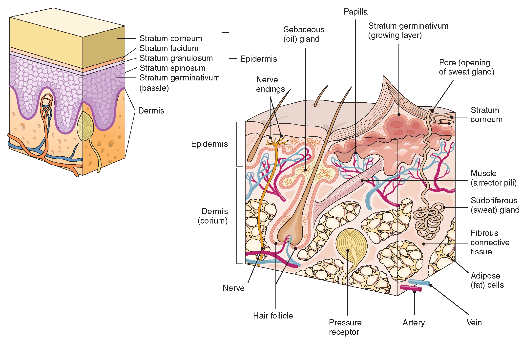
The Epidermis The epidermis is composed of keratinized, stratified squamous epithelium. It is made of four or five layers of epithelial cells, depending on its location in the body. It does not have any blood vessels within it (i.e., it is avascular). Skin that has four layers of cells is referred to as "thin skin."
epidermis structure systems Biological Science Picture Directory
Skin also helps maintain a constant body temperature. Human skin is only about 0.07 inches (2 mm) thick. Skin is made up of two layers that cover a third fatty layer. The outer layer is called the epidermis; it is a tough protective layer that contains melanin (which protects against the rays of the sun and gives the skin its color).

The Structure Of Human Skin Cells Stock Illustration Download Image Now Hair Follicle
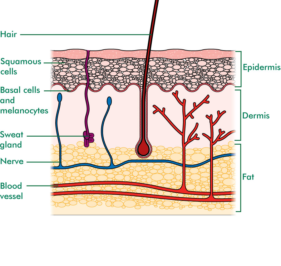
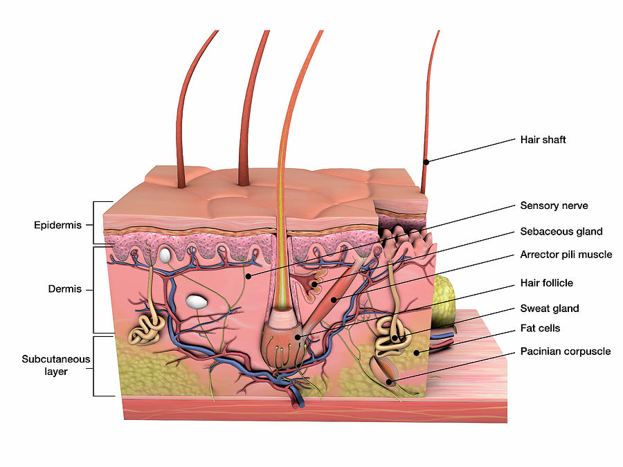
This diagram shows the layers found in skin. There are three main layers: the epidermis, dermis and hypodermis. There are also sweat glands, and hairs, which have sebaceous glands, and a smooth muscle called the arrector pili muscle, associated with them.
Anatomy Of Human Skin With Labels Photograph by Hank Grebe Pixels
Figure 1. The skin is composed of two main layers: the epidermis, made of closely packed epithelial cells, and the dermis, made of dense, irregular connective tissue that houses blood vessels, hair follicles, sweat glands, and other structures. Beneath the dermis lies the hypodermis, which is composed mainly of loose connective and fatty tissues.
The Integumentary System (Structure and Function) (Nursing) Part 1
Functions of the skin. Some of the many roles of skin include: Protecting against pathogens. Langerhans cells in the skin are part of the immune system. Storing lipids (fats) and water. Creating.
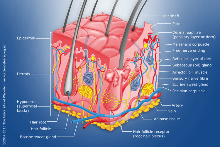
Diagram of human skin structure — Science Learning Hub
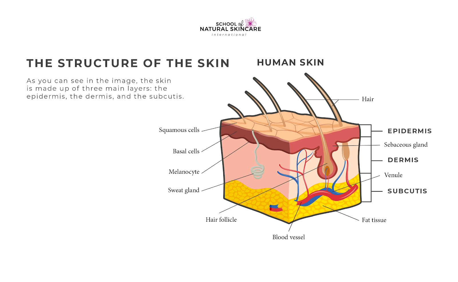
What is skin? Although you may not realise it, your skin is your largest organ. Learn more about its parts, how it functions and how to keep it healthy. Parts of the skin Skin covers your body and has three layers: The top layer is the epidermis (outer layer). This is a thin layer. It provides a waterproof barrier for your body.

Human skin diagram Subcutaneous tissue, Skin structure, Epidermis
The skin is the body's largest and primary protective organ, covering its entire external surface and serving as a first-order physical barrier against the environment. Its functions include temperature regulation and protection against ultraviolet (UV) light, trauma, pathogens, microorganisms, and toxins.

Skin Structure infographic LifeMap Discovery
1/3 Synonyms: none This article will describe the anatomy and histology of the skin. Undoubtedly, the skin is the largest organ in the human body; literally covering you from head to toe. The organ constitutes almost 8-20% of body mass and has a surface area of approximately 1.6 to 1.8 m2, in an adult.

Dermatology Diagram Show Human Skin Structure Stock Illustration Download Image Now Anatomy
Explore Skin Diagram with BYJU'S. Diagram of the skin is illustrated in detail with neat and clear labelling. Also available for free download

Skin Definition, Structure And Functions Of Skin
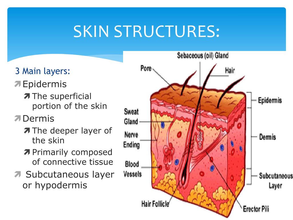
Skin is part of the integumentary system and considered to be the largest organ of the human body. There are three main layers of skin: the epidermis, the dermis, and the hypodermis (subcutaneous fat). The focus of this topic is on the epidermal and dermal layers of skin. Skin appendages such as sweat glands, hair follicles, and sebaceous glands are reviewed in-depth elsewhere.[1]

loadBinary_006.gif (992×779) Skin anatomy, Integumentary system, Human integumentary system
Figure 1. Layers of Skin. The skin is composed of two main layers: the epidermis, made of closely packed epithelial cells, and the dermis, made of dense, irregular connective tissue that houses blood vessels, hair follicles, sweat glands, and other structures.
Skin diagram to label Labelled diagram
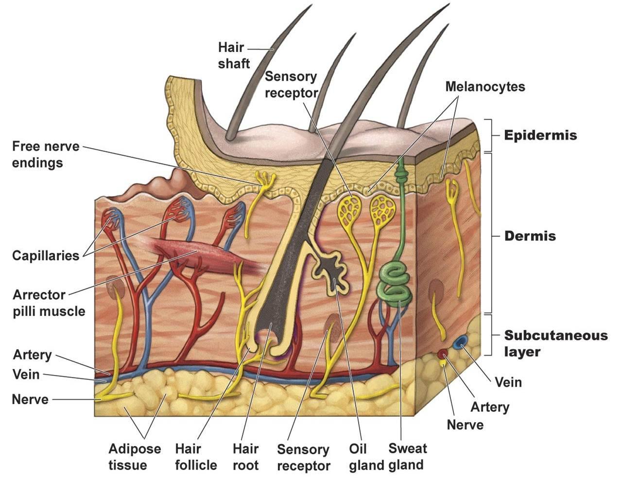
Key facts about the integumentary system; Skin: Functions: chemical and mechanical barrier, biosynthesis, control of body temperature, sensory Layers: Epidermis (Stratum Basale, Spinosum, Granulosum, Lucidum, Corneum) and dermis (papillary, reticular) Mnemonic: British and Spanish Grannies Love Cornflakes Hair: Types: vellus and terminal Structure: Follicle and bulb (shaft, inner root sheath.